20k gallen bladder tear test|blood test for gallbladder inflammation : ODM A hepatobiliary iminodiacetic acid (HIDA) scan is an imaging procedure used to . A plant tissue culture medium containing glucose or fructose sterilized by autoclaving inhibits the growth of carrot root tissue cultures. While some inhibition occurs when the sugar alone is .
{plog:ftitle_list}
Step 4: Transfer the solution to an autoclavable bottle. Sterilize the solution by autoclaving (20 minutes at 15 lb/sq.in. (psi) from 121-124°C on liquid cycle). STORAGE Solution can be stored .


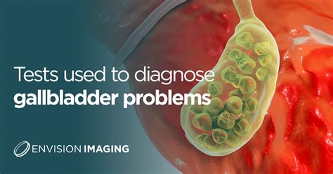
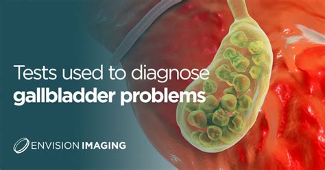
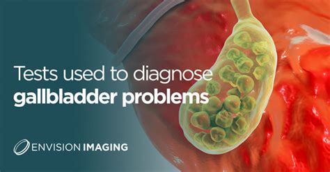
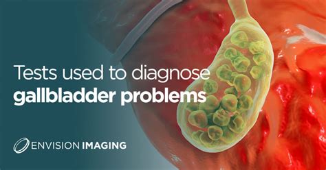
Gallbladder problems are diagnosed through various tests. These may include: Liver tests, which are blood tests that can show evidence of gallbladder disease. A check of the blood's amylase.HIDA scan for gallbladder: This test uses a radioactive compound to “trace” the path .
urine tests for gallbladder problems
Lipase test: Lipase is a protein that helps your body absorb fats. Your doctor can .MD's Digestive Disorders reference library for patients interested in finding . A hepatobiliary iminodiacetic acid (HIDA) scan is an imaging procedure used to . Diagnostic tests like CT Scan, Urine & Blood Tests, X-rays and an Ultrasound .

Gallbladder problems are diagnosed through various tests. These may include: Liver tests, which are blood tests that can show evidence of gallbladder disease. A check of the blood's amylase.
The gallbladder releases bile into the small bowel to help break down fats. A gallbladder rupture is a medical condition where the gallbladder wall leaks or bursts. Ruptures are commonly. A hepatobiliary iminodiacetic acid (HIDA) scan is an imaging procedure used to diagnose problems of the liver, gallbladder and bile ducts. For a HIDA scan, also known as cholescintigraphy or hepatobiliary scintigraphy, a radioactive tracer . Diagnostic tests like CT Scan, Urine & Blood Tests, X-rays and an Ultrasound can help determine gallbladder disease. The tests typically include: Complete blood count (CBC), which can detect increased white blood cells in people with gallbladder inflammation; Liver function tests (LFTs), which can detect increased liver enzymes when a gallstone blocks the bile duct
This condition, also known as gallbladder perforation, can lead to severe complications, including infection and peritonitis, if not treated promptly. In this article, we’ll explore the causes, symptoms, diagnosis, and treatment options for a ruptured gallbladder. An abdominal ultrasound is an imaging test using sound waves that can used to view internal organs, including the gallbladder. A healthcare provider may order one if symptoms, such as abdominal pain, nausea, vomiting, and chills, suggest a problem with the gallbladder. A gallbladder ultrasound is noninvasive and painless.
Here are the primary tests used: Physical examination: The doctor may check for pain in the abdomen, particularly in the upper right side, which could indicate gallbladder problems. Blood. A gallbladder radionuclide scan is an imaging test that uses radiation to detect: infection. disease. bile fluid leakage. blockage in your gallbladder. The procedure uses radioactive. Bladder rupture can be diagnosed through a combination of physical examination, medical history, and imaging tests such as: Abdominal ultrasound; Computed tomography (CT) scan; Magnetic resonance imaging (MRI) Cystogram (X-ray of the bladder) Treatment: Treatment for bladder rupture depends on the severity of the injury.
Gallbladder problems are diagnosed through various tests. These may include: Liver tests, which are blood tests that can show evidence of gallbladder disease. A check of the blood's amylase. The gallbladder releases bile into the small bowel to help break down fats. A gallbladder rupture is a medical condition where the gallbladder wall leaks or bursts. Ruptures are commonly. A hepatobiliary iminodiacetic acid (HIDA) scan is an imaging procedure used to diagnose problems of the liver, gallbladder and bile ducts. For a HIDA scan, also known as cholescintigraphy or hepatobiliary scintigraphy, a radioactive tracer . Diagnostic tests like CT Scan, Urine & Blood Tests, X-rays and an Ultrasound can help determine gallbladder disease.
The tests typically include: Complete blood count (CBC), which can detect increased white blood cells in people with gallbladder inflammation; Liver function tests (LFTs), which can detect increased liver enzymes when a gallstone blocks the bile duct This condition, also known as gallbladder perforation, can lead to severe complications, including infection and peritonitis, if not treated promptly. In this article, we’ll explore the causes, symptoms, diagnosis, and treatment options for a ruptured gallbladder. An abdominal ultrasound is an imaging test using sound waves that can used to view internal organs, including the gallbladder. A healthcare provider may order one if symptoms, such as abdominal pain, nausea, vomiting, and chills, suggest a problem with the gallbladder. A gallbladder ultrasound is noninvasive and painless. Here are the primary tests used: Physical examination: The doctor may check for pain in the abdomen, particularly in the upper right side, which could indicate gallbladder problems. Blood.
A gallbladder radionuclide scan is an imaging test that uses radiation to detect: infection. disease. bile fluid leakage. blockage in your gallbladder. The procedure uses radioactive.
gallbladder x ray blood test

adiponectin elisa kit r&
gallbladder test results
G.M.Kamalakannan, Principal Scientist and Deputy Head, CSMST, CSIR-NAL Autoclave has enabled the replacement of metallic aerospace structures by advanced composites structures.
20k gallen bladder tear test|blood test for gallbladder inflammation