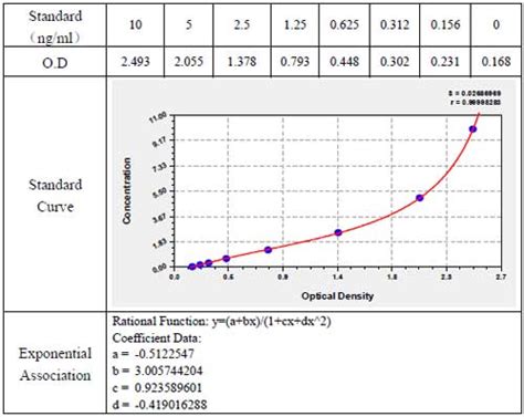
elisa test graph|how to interpret elisa data : private label ELISA data analysis in EXCELEliza assay, construct, standard curve, concentration, unknown, samples, raw data, result, bar chart, disease, control, well, dup. For organisations outside the UK, please contact your national dealer who can be .
{plog:ftitle_list}
As the autoclaving of a sugar together with other nutrient components enhances its degradation with the associated formation of toxic products, it is advisable to autoclave it separately from other medium components.What is the clinical effectiveness of steam sterilization of non-sterile gauze in an operating room pack? What are the harms associated with placing non-sterile gauze in an operating room pack, to be steam sterilized?
Add Unknowns to your Graph. Prism's automatic graph includes the data from the data sheet and the curve. To add the "unknowns" to the graph: Switch to the Graphs section of your project. Click on the Change button and then select Data on Graph. The dialog box shows all data and results tables that are represented on the graph. Click on the Add . ELISA data analysis in EXCELEliza assay, construct, standard curve, concentration, unknown, samples, raw data, result, bar chart, disease, control, well, dup.An example of a competition ELISA to test for antigen based on the direct detection method is shown in Figure 5 . Remove liquid and wash plate Remove liquid and wash plate Remove liquid and wash plate Remove liquid and wash plate In this example the antigen concentration in the sample was low. The

The Enzyme-Linked Immunosorbent Assay (ELISA) is a highly sensitive procedure to quantify the concentration of an antibody or antigen in a sample. The estimation of the analyte concentration depends upon the construction of a standard curve. The standard curve is prepared by making serial dilutions of one known concentration of the analyte .
ELISA test results, what does a positive ELISA test tell you? ELISA results may be interpreted quantitatively, qualitatively or semi-quantitatively. In a quantitative assay, a serial dilution of a known standard is used to enable the generation of a standard curve, normally of optical density (OD) versus concentration. From this, the precise . Increasing the binding affinity of an antibody to its target antigen is a crucial task in antibody therapeutics development. This paper presents a pretrainable geometric graph neural network .
Researchers consider ELISA to be the gold standard of immunoassays. Tests that use ELISA can help diagnose a wide range of conditions, from bacterial and viral infections (like Lyme disease and HIV) to endocrine conditions, like thyroid disease.. Home pregnancy tests are even based on the ELISA technique. They detect the presence of a hormone called human chorionic .
Before running an ELISA, consider the following best practices to get accurate and consistent data: 1. Run samples in replicate. To help evaluate the extent of error, each standard and sample should be tested in replicate (duplicate or triplicate, depending on the number of samples and room on the plate). The final report document generated in ELISA Tool contains: results of the absorbance measurement for the calibration standards, selected approximation (curve fitting) model, absorbance results for the tested samples, respective concentrations in the tested samples calculated from the calibration curve, and corresponding graphs with a linear .points in the graph (we suggest that a suitable computer program be used for this). We recommend including a standard on each ELISA plate to provide a standard curve for each plate used. A representative standard curve is shown in the figure below, from human HIF1 alpha SimpleStep ELISATM kit (ab171577). Each point on the graph represents the .
If your software allows it, 4-PL and 5-PL will fit most ELISA calibration standard curves. If not, the best option is to use a semi-log or a log/log plot. Competitive ELISA standard curve. For competitive ELISA, the antigen concentration is determined from the standard curve in the same manner as a conventional ELISA.Variations between ELISA protocols A. Antigen Immobilization Antigen immobilization varies between two principle techniques. In a traditional (direct coating) ELISA, antigens are directly attached to the plate by passive adsorption, usually using a carbonate/bicarbonate buffer at pH >9. Most but not all proteinsELISA is a popular technique for research and diagnostic samples and can be used as both a single sample test or high throughput method for screening. PathScan® Phospho-Akt (Thr308) Sandwich ELISA Kit #7252: The relationship between protein concentration of lysates from untreated and PDGF-treated NIH/3T3 cells and kit assay optical density .
Learn how to perform ELISA data analysis. Get to know the different aspects to consider for more consistent and accurate ELISA data. Furthermore, we provide a step-by-step guide to create a standard curve for analysis.The sensitivity of this ELISA test is 0.05 µIU/mL. The Thyroid Stimulating Hormone (TSH) ELISAis intended for the quantitative measurement of TSH in human serum. It is a solid phase sandwich ELISA . Graph paper Precautions and Disclaimer This product is for R&D use only, not for drug, household, or other uses. Please consult the Safety ELISA: 1. Record results of Widal test using + or - signs: extremely agglutinated (+++) very agglutinated (++) a little agglutinated (+) . and hormones using antibodies and color changes. ELISA is a common medical and research lab technique. This page titled Lab 13: ELISA is shared under a CC BY-SA license and was authored, remixed, and/or .
The antibodies utilized for an ELISA test can be monoclonal or polyclonal. ELISA results yield three different types of data: • Quantitative: With quantitative data, . Any known concentrations of antigen are utilized to give the standard curve on the graph. Then, that data can be used to measure the concentration of the unknown samples when .Prism's automatic graph includes the data from the Data sheet and the curve. To add the "unknowns" to the graph: Switch to the graph Transform of Data 1 graph. Choose Change. Add Data Sets.. From the top drop-down box in the Add Data Sets to Graph dialog, select the data set containing the .Interpolated X values.
An ELISA is typically performed in a plastic microstrip plate that has 12 wells. Each strip has wells for a positive control, a negative . PCR (polymerase chain reaction). Using the graph, explain when a viral PCR test would be most useful. Questions — ELISA Design 3. Refer to the diagram, and notice where the primary antibody binds antigen .As to better illustrate how ELISA test works, this section took a deeper insight into: a definition of the 'sensitivity', standard curve calculation, and control samples in ELISA test, followed by a list of diseases in which ELISA test could be applied. . From the X axis of the standard curve graph, extend a horizontal line from this .Generally, a sandwich ELISA test includes the following reaction steps: immobilization of capture antigen/antibody on a microplate, adding buffer solution, detection of antibody and several steps of washing and incubation, addition of 3,3′,5,5′-tetramethylbenzidine (TMB) and stopping solutions. . the correlation graph between the .Draw a best-fit curve through the points in the graph (we suggest that a suitable computer program be used for this). We recommend including a standard on each ELISA plate to provide a standard curve for each plate used. A representative standard curve is shown in the figure below from the human HIF1 alpha SimpleStep ELISA kit® (ab171577 .
A Competitive ELISA, also known as an inhibition assay, is an ELISA that can measure the concentration of an analyte through its interference in the ELISA assay signal. Competitive ELISAs are great at detecting small analytes such as lipids and can detect these analytes in complex mixtures like plasma, serum, or cell extracts. These assays . This video supports BABEC's "Exploring Vaccines with ELISA" lab.Register for a free account at https://babec.org/ to download this lesson and more!3. View the graph. The graph Prism makes automatically is fairly complete. You can customize the symbols, colors, axis labels, etc. You can also choose to plot the individual duplicates rather than plot the means. Since the unknowns have no X value, they are not included on the graph. 4. Choose the standard curve analysis
How to use Microsoft Excel to quantify ELISA data. The Enzyme Linked Immunosorbent Assay . You can try different regions of the linear end of the graph looking for the R 2 value closest to 1. So to translate the OD405 value in cell B1 of the ELISA well into ng/ml values, you can simple put the equation “=((B1*3.1685)-0.02563)”. .SimpleStep ELISA ® kits drastically reduce assay times without compromising on performance. We compared the sensitivity of SimpleStep ELISA® kits to the most popular competitor brand of ELISA kits for 75 popular human and mouse proteins . SimpleStep ELISA ® kits show superior sensitivity in 56 out of 75 cases.
how to interpret elisa results
how to interpret elisa data
how to graph elisa results
I’m designing a part that will need to be autoclaved—it will be under steam at 121°C for about 15 min per job and I will want it to be able to go through the autoclave repeatedly.
elisa test graph|how to interpret elisa data