test for patellar tendon tear|patellar tendon enthesopathy : OEM A traumatic rupture of the patellar tendon caused by a tension overload during activity in a patient at risk. Diagnosis can be confirmed by physical exam and radiographs for complete tears. Partial tears may need an MRI to confirm the diagnosis. An authorized training session must be successfully completed prior to use of autoclaves. Heat-insulating gloves that provide complete coverage of hands and forearms must be worn when .
{plog:ftitle_list}
Biological spore testing is performed monthly on all research autoclaves used .
A traumatic rupture of the patellar tendon caused by a tension overload during activity in a patient at risk. Diagnosis can be confirmed by physical exam and radiographs for complete tears. Partial tears may need an MRI to confirm the diagnosis.Diagnosis is primarily made clinically with tenderness to palpation at the distal pole .
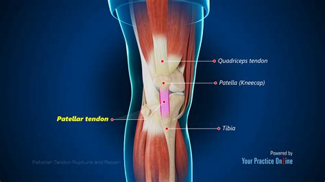
A traumatic rupture of the patellar tendon caused by a tension overload during activity in a patient at risk. Diagnosis can be confirmed by physical exam and radiographs for complete tears. Partial tears may need an MRI to confirm the diagnosis. A patellar tendon tear is the partial or complete rupture of the band of connective tissue that attaches the kneecap to the shin bone. When this happens, you may be unable to walk or fully straighten the leg. Knee pain, bruising, and swelling are common symptoms.
While this part of the examination can be painful, it is important to identify a patellar tendon tear. Imaging Tests. To confirm the diagnosis, your doctor may order some imaging tests, such as an X-ray or magnetic resonance imaging (MRI) scan. X-rays. The kneecap moves out of place when the patellar tendon tears. Ultrasound. This test uses sound waves to create an image of your knee, revealing tears in your patellar tendon. Magnetic resonance imaging (MRI). MRI uses a magnetic field and radio waves to create detailed images that can reveal subtle changes in the patellar tendon.
Diagnosis of patella tendon tear should be made as early as possible to avoid poor functional outcome with a loss of full knee flexion and decreased quadriceps strength. Accurate diagnosis depends on detailed history, physical examination and radiographic examinations. Range of motion (ROM) testing and muscle strength testing are essential aspects of the knee exam, especially in the setting of a suspected patellar tendon rupture. Patients with an acute patellar tendon rupture will have decreased ROM of the knee due to pain and disruption of the extensor mechanism.
Diagnosis is primarily made clinically with tenderness to palpation at the distal pole of patella in full extension. Treatment is generally nonoperative with resting, ice, activity modifications and physical therapy to focus on hamstring, quadriceps and core strengthening.
September 19, 2023. A patella tendon rupture or patella tendon strain is a partial, or complete tear of the patella tendon at the front of the knee. Advert. Medically reviewed by Dr Chaminda Goonetilleke, 2nd Jan. 2022. Symptoms. Patella tendon ruptures are extremely painful. Signs and symptoms include: Severe pain in the front of the knee. If you’ve torn your patellar tendon, you may be asked to have some scans to confirm the diagnosis and work out the extent of the damage – an x-ray to check the bone and / or an MRI scan to get a better view of the tendon tissues. Could a partial patellar tendon tear heal on its own? Yes, it’s possible. Ultrasound. Best imaged with longitudinal scans using high-frequency linear transducers; the tendon normally appears as a continuous well-defined hyperechoic fibrillar structure bridging the patella and the tibial tuberosity while tears usually appear as hypoechoic areas of interruption of the fibrillar pattern 3. Treatment and prognosis. A traumatic rupture of the patellar tendon caused by a tension overload during activity in a patient at risk. Diagnosis can be confirmed by physical exam and radiographs for complete tears. Partial tears may need an MRI to confirm the diagnosis.
A patellar tendon tear is the partial or complete rupture of the band of connective tissue that attaches the kneecap to the shin bone. When this happens, you may be unable to walk or fully straighten the leg. Knee pain, bruising, and swelling are common symptoms.

describe autoclaving process
what is a patella evaluation

While this part of the examination can be painful, it is important to identify a patellar tendon tear. Imaging Tests. To confirm the diagnosis, your doctor may order some imaging tests, such as an X-ray or magnetic resonance imaging (MRI) scan. X-rays. The kneecap moves out of place when the patellar tendon tears. Ultrasound. This test uses sound waves to create an image of your knee, revealing tears in your patellar tendon. Magnetic resonance imaging (MRI). MRI uses a magnetic field and radio waves to create detailed images that can reveal subtle changes in the patellar tendon.
Diagnosis of patella tendon tear should be made as early as possible to avoid poor functional outcome with a loss of full knee flexion and decreased quadriceps strength. Accurate diagnosis depends on detailed history, physical examination and radiographic examinations. Range of motion (ROM) testing and muscle strength testing are essential aspects of the knee exam, especially in the setting of a suspected patellar tendon rupture. Patients with an acute patellar tendon rupture will have decreased ROM of the knee due to pain and disruption of the extensor mechanism. Diagnosis is primarily made clinically with tenderness to palpation at the distal pole of patella in full extension. Treatment is generally nonoperative with resting, ice, activity modifications and physical therapy to focus on hamstring, quadriceps and core strengthening.
September 19, 2023. A patella tendon rupture or patella tendon strain is a partial, or complete tear of the patella tendon at the front of the knee. Advert. Medically reviewed by Dr Chaminda Goonetilleke, 2nd Jan. 2022. Symptoms. Patella tendon ruptures are extremely painful. Signs and symptoms include: Severe pain in the front of the knee.
If you’ve torn your patellar tendon, you may be asked to have some scans to confirm the diagnosis and work out the extent of the damage – an x-ray to check the bone and / or an MRI scan to get a better view of the tendon tissues. Could a partial patellar tendon tear heal on its own? Yes, it’s possible.
torn patellar tendon symptoms
describe the use of an autoclave in microbiology laboratory

Sign In - Siltex Online Database. Ultrasonic Cleaners. Soniclean is one of the .
test for patellar tendon tear|patellar tendon enthesopathy