acl tear orthopedic tests|acl tests physical exam : warehouse These tests may include: X-rays. X-rays may be needed to rule out a bone fracture. However, X-rays don't show soft tissues, such as ligaments and tendons. Magnetic resonance imaging (MRI). An MRI uses radio waves and a strong magnetic field to create images of both hard and soft tissues in your body. Autoclave Sterilization: Autoclaves provide a physical method for disinfection and sterilization. They work with a combination of steam, pressure and time. Autoclaves operate at .
{plog:ftitle_list}
C. Manual Cleaning Most instrument manufacturers recommend ultrasonic cleaning as the best and most effective way to clean surgical instruments, particularly those with hinges, locks and .
ACL tears are common athletic injuries leading to anterior and lateral rotatory instability of the knee. Diagnosis can be suspected clinically with presence of a traumatic knee effusion with increased laxity on Lachman's test but requires MRI studies to confirm diagnosis.Posterolateral corner (PLC) injuries are traumatic knee injuries that are associated with later.
ACL tears are common athletic injuries leading to anterior and lateral rotatory instability of the knee. Diagnosis can be suspected clinically with presence of a traumatic knee effusion with increased laxity on Lachman's test but requires MRI studies to confirm diagnosis. These tests may include: X-rays. X-rays may be needed to rule out a bone fracture. However, X-rays don't show soft tissues, such as ligaments and tendons. Magnetic resonance imaging (MRI). An MRI uses radio waves and a strong magnetic field to create images of both hard and soft tissues in your body.
The anterior drawer test is a physical examination doctors use to test the stability of the knee’s anterior cruciate ligament (ACL). Doctors may use this test, along with images and other.Imaging Tests. Other tests which may help your doctor confirm your diagnosis include: X-rays. Although they will not show any injury to your anterior cruciate ligament, X-rays can show whether the injury is associated with a broken bone. Magnetic resonance imaging (MRI) scan.
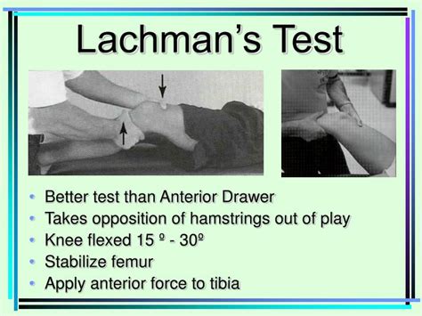
An ACL tear is an injury to the anterior cruciate ligament (ACL) in your knee. The recovery time is usually six to nine months after surgery. The Lachman test is done to check for an anterior cruciate ligament (ACL) injury or tea r. The ACL connects two of the three bones that form your knee joint: patella, or kneecap. femur,.
The Lachman test is the most accurate test for detecting an ACL tear. Magnetic resonance imaging is the primary study used to diagnose ACL injury in the United States. It can also identify.
An ACL injury is a tear or sprain of the anterior cruciate ligament (ACL) — one of the major ligaments in your knee. Ligaments are strong bands of tissue that connect one bone to another. The ACL, one of two ligaments that cross in the middle of the knee connecting the thigh bone (femur) to the shinbone (tibia), help stabilize the joint. An ACL injury is a tear or sprain of the anterior cruciate (KROO-she-ate) ligament (ACL) — one of the strong bands of tissue that help connect your thigh bone (femur) to your shinbone (tibia). ACL injuries most commonly occur during sports that involve sudden stops or changes in direction, jumping and landing — such as soccer, basketball . In the anterior drawer test, the examiner moves the tibia forward with respect to the femur, with the patient’s knee at 90 degrees of flexion and the feet flat; excessive anterior. ACL tears are common athletic injuries leading to anterior and lateral rotatory instability of the knee. Diagnosis can be suspected clinically with presence of a traumatic knee effusion with increased laxity on Lachman's test but requires MRI studies to confirm diagnosis.
These tests may include: X-rays. X-rays may be needed to rule out a bone fracture. However, X-rays don't show soft tissues, such as ligaments and tendons. Magnetic resonance imaging (MRI). An MRI uses radio waves and a strong magnetic field to create images of both hard and soft tissues in your body.

is pipette reef safe
tests to determine acl tear

The anterior drawer test is a physical examination doctors use to test the stability of the knee’s anterior cruciate ligament (ACL). Doctors may use this test, along with images and other.Imaging Tests. Other tests which may help your doctor confirm your diagnosis include: X-rays. Although they will not show any injury to your anterior cruciate ligament, X-rays can show whether the injury is associated with a broken bone. Magnetic resonance imaging (MRI) scan.
An ACL tear is an injury to the anterior cruciate ligament (ACL) in your knee. The recovery time is usually six to nine months after surgery. The Lachman test is done to check for an anterior cruciate ligament (ACL) injury or tea r. The ACL connects two of the three bones that form your knee joint: patella, or kneecap. femur,. The Lachman test is the most accurate test for detecting an ACL tear. Magnetic resonance imaging is the primary study used to diagnose ACL injury in the United States. It can also identify.
An ACL injury is a tear or sprain of the anterior cruciate ligament (ACL) — one of the major ligaments in your knee. Ligaments are strong bands of tissue that connect one bone to another. The ACL, one of two ligaments that cross in the middle of the knee connecting the thigh bone (femur) to the shinbone (tibia), help stabilize the joint.
An ACL injury is a tear or sprain of the anterior cruciate (KROO-she-ate) ligament (ACL) — one of the strong bands of tissue that help connect your thigh bone (femur) to your shinbone (tibia). ACL injuries most commonly occur during sports that involve sudden stops or changes in direction, jumping and landing — such as soccer, basketball .
self test acl tear
is pipette safe for babies

NOTE: The main handle and bottom cap components of handles marked “AUTOCLAVE” are compatible with the autoclave methods identified which are provided for facilities who wish to .
acl tear orthopedic tests|acl tests physical exam