ankle lateral ligament tear test|lateral x ray ankle ligaments : traders Ankle sprains are one of the most common musculoskeletal and sports-related injuries, constituting nearly 25% of all musculoskeletal trauma cases and almost 40% of all sports-related trauma cases. 40%-50% of these case have reported to have long term residual symptoms with almost 20% of acute ankle . See more Researchers writing in the current issue of the journal Science report that the organism, dubbed Strain 121, can survive at temperatures of 121 degrees Celsius or 250 .
{plog:ftitle_list}
$869.99
Ankle sprains are one of the most common musculoskeletal and sports-related injuries, constituting nearly 25% of all musculoskeletal trauma cases and almost 40% of all sports-related trauma cases. 40%-50% of these case have reported to have long term residual symptoms with almost 20% of acute ankle . See moreLigaments of the ankle Lateral ankle ligaments The lateral side of the anklehas three supporting ligaments: the anterior talofibular ligament (ATFL), the posterior talofibular ligament . See more
1. Anterior Drawer Test It is used to assess the integrity of the ATFL based on the anterior translation of the talus under the tibia in a sagittal plane. Procedure: The patient is in . See more

1. Sensitivity values for the Anterior Drawer test have been shown to be between 32% to 80% while specificity value has been . See moreAn anterior drawer is done to test the integrity of the ATFL and CFL. With the ankle in plantarflexion, the heel is grasped with the tibia stabilised and drawn anteriorly. Talar tilt is done to assess the integrity of the ATFL and CFL .
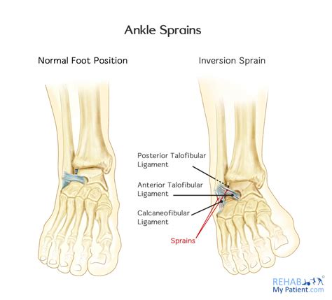
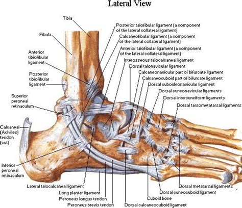
The lateral side of the ankle has three supporting ligaments: the anterior talofibular ligament (ATFL), the posterior talofibular ligament (PTFL) and the calcaneofibular ligament(CFL). The three ligaments are together called the Lateral Collateral Ligament Complex.An anterior drawer is done to test the integrity of the ATFL and CFL. With the ankle in plantarflexion, the heel is grasped with the tibia stabilised and drawn anteriorly. Talar tilt is done to assess the integrity of the ATFL and CFL laterally and the deltoid ligament medially.
lateral x ray ankle ligaments
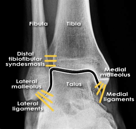
forward shift of more than 8 mm on a lateral radiograph is considered diagnostic for an ATFL tear

Objective Assessment: Assessment of ligamentous laxity: Swelling. Isometric ankle eversion/abduction. Chronic Ankle Instability Checklist. Following LAS, a comprehensive assessment of the factors contributing factors can help predict the prognosis. The following is a set of markers that are associated with CAI: ROM Markers:For management purposes, ankle injuries can be considered under the following headings: • Mild to moderate lateral ligament complex sprain – treatable with early mobilisation and guided proprioceptive and strengthening rehabilitation program • Severe lateral ligament complex sprain – immobilisation for a
This protocol is intended to guide clinicians through non-operative management of lateral ankle sprain. This protocol is time based (dependent on tissue healing) as well as criterion based.
Patients with ankle sprains (stretching, partial rupture, or complete rupture of at least one ligament) constitute a large percentage of these injuries. The epidemiology, presentation, and evaluation of common ankle sprains are reviewed here.
lateral ligament ankle injury
• Complete tear of the anterior talofibular ligament, the calcaneofibular, and the posterior talofibular ligament. • Significant tenderness and swelling around the ankle.
The Ottawa ankle rules are used to rule out a fracture, and radiographs are indicated if there is pain within the malleolar zone that is accompanied by any of the following findings: (1) tenderness along the tip of the posterior edge of the lateral malleolus, (2) tenderness over the medial malleolus, and/or (3) the inability of the patient to be.Two of the most common manual stress tests for the ankle are the anterior drawer and talar tilt tests. The anterior drawer test is performed with the application of an anterior load applied to the ankle. . Hofmann O.E. Biomechanical comparison of reconstruction techniques in simulated lateral ankle ligament injury. Am J Sports Med. 1995;23(6 .
The lateral side of the ankle has three supporting ligaments: the anterior talofibular ligament (ATFL), the posterior talofibular ligament (PTFL) and the calcaneofibular ligament(CFL). The three ligaments are together called the Lateral Collateral Ligament Complex.An anterior drawer is done to test the integrity of the ATFL and CFL. With the ankle in plantarflexion, the heel is grasped with the tibia stabilised and drawn anteriorly. Talar tilt is done to assess the integrity of the ATFL and CFL laterally and the deltoid ligament medially.
forward shift of more than 8 mm on a lateral radiograph is considered diagnostic for an ATFL tearObjective Assessment: Assessment of ligamentous laxity: Swelling. Isometric ankle eversion/abduction. Chronic Ankle Instability Checklist. Following LAS, a comprehensive assessment of the factors contributing factors can help predict the prognosis. The following is a set of markers that are associated with CAI: ROM Markers:
For management purposes, ankle injuries can be considered under the following headings: • Mild to moderate lateral ligament complex sprain – treatable with early mobilisation and guided proprioceptive and strengthening rehabilitation program • Severe lateral ligament complex sprain – immobilisation for aThis protocol is intended to guide clinicians through non-operative management of lateral ankle sprain. This protocol is time based (dependent on tissue healing) as well as criterion based. Patients with ankle sprains (stretching, partial rupture, or complete rupture of at least one ligament) constitute a large percentage of these injuries. The epidemiology, presentation, and evaluation of common ankle sprains are reviewed here.• Complete tear of the anterior talofibular ligament, the calcaneofibular, and the posterior talofibular ligament. • Significant tenderness and swelling around the ankle.
lateral ligament ankle anatomy
The Ottawa ankle rules are used to rule out a fracture, and radiographs are indicated if there is pain within the malleolar zone that is accompanied by any of the following findings: (1) tenderness along the tip of the posterior edge of the lateral malleolus, (2) tenderness over the medial malleolus, and/or (3) the inability of the patient to be.

how to use a refractometer aquarium
how to use a refractometer battery charge
Test cycles Bowie and Dick/Helix/Vacuum test Chemical indicator in a product challenge device Periodic testing as referred to in ISO 17665-1
ankle lateral ligament tear test|lateral x ray ankle ligaments