acl mcl tear test|acl vs mcl tear symptoms : China An MRI can show the extent of an ACL injury and signs of damage to other tissues in the knee, including the cartilage. Ultrasound. Using sound waves to visualize internal structures, ultrasound may be used to check for injuries in the ligaments, tendons and muscles of . Le traitement classe II ou III : il offre une protection intermédiaire entre l'autoclave et le bois brut pour un usage en extérieur. Le traitement fongicide et/ou insecticide : son application est .
{plog:ftitle_list}
The report analyses the key growth drivers, opportunities, and challenges influencing the global steam autoclave market.
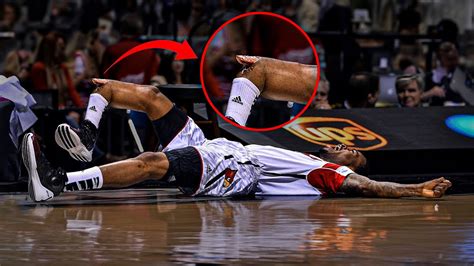
worst NBA injuries acl mcl
ACL vs. MCL tears: Although symptoms of ACL and MCL tears are similar, a few key differences will help identify whether the injury affected the ACL or MCL. An ACL tear will . Anterior Cruciate Ligament (ACL) Lacchman’s test. It is performed with the patient supine and the knee flexed 20–30°. The examiner grasps the distal femur (from lateral side) with one hand and the proximal tibia with the other hand (from medial side). The lower leg is given a brisk forward tug in an attempt to identify a discrete endpoint.
ACL vs. MCL tears: Although symptoms of ACL and MCL tears are similar, a few key differences will help identify whether the injury affected the ACL or MCL. An ACL tear will have a more distinctive and loud popping sound than an MCL tear.
is the special points test hard
An MRI can show the extent of an ACL injury and signs of damage to other tissues in the knee, including the cartilage. Ultrasound. Using sound waves to visualize internal structures, ultrasound may be used to check for injuries in the ligaments, tendons and muscles of . An MCL tear is damage to the medial collateral ligament, which is a major ligament that’s located on the inner side of your knee. The tear can be partial (some fibers in the ligament are torn) or complete (the ligament is torn into two pieces). ACL tears are common athletic injuries leading to anterior and lateral rotatory instability of the knee. Diagnosis can be suspected clinically with presence of a traumatic knee effusion with increased laxity on Lachman's test but requires MRI studies to confirm diagnosis. What causes ACL tears? Anything that puts too much force on your knee can tear your ACL. ACL tears happen when your knee moves or twists more than it naturally can. The most common causes of ACL tears include: Sports injuries. Car accidents. Falls. ACL tear risk factors. Anyone can experience an ACL tear.
A medial collateral ligament (MCL) injury is a stretch, partial tear, or complete tear of the ligament on the inside of the knee. A valgus trauma or external tibia rotation are the causes of this injury. A medial collateral ligament (MCL) knee injury is a traumatic knee injury that typically occurs as a result of a sudden valgus force to the lateral aspect of the knee. Diagnosis can be suspected with increased valgus laxity on physical exam but requires MRI for confirmation. Treatment is generally nonoperative with bracing.
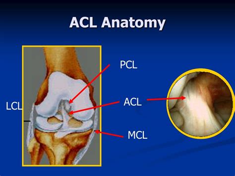
Two of the most frequently encountered knee injuries are tears of the anterior cruciate ligament (ACL) and the medial collateral ligament (MCL). While both injuries involve ligament damage and can result in pain and instability, they differ in their causes, symptoms, and treatment approaches. anterior cruciate ligament. ( ACL. ), posterior cruciate ligament. ( PCL. ), medial collateral ligament. ( MCL. ), and. lateral collateral ligament. ( LCL. ) result in knee pain and instability. Various maneuvers can be used to evaluate the stability of the joint and usually suffice to diagnose collateral. ligament. tears. An. MRI. is the best.
Anterior Cruciate Ligament (ACL) Lacchman’s test. It is performed with the patient supine and the knee flexed 20–30°. The examiner grasps the distal femur (from lateral side) with one hand and the proximal tibia with the other hand (from medial side). The lower leg is given a brisk forward tug in an attempt to identify a discrete endpoint. ACL vs. MCL tears: Although symptoms of ACL and MCL tears are similar, a few key differences will help identify whether the injury affected the ACL or MCL. An ACL tear will have a more distinctive and loud popping sound than an MCL tear. An MRI can show the extent of an ACL injury and signs of damage to other tissues in the knee, including the cartilage. Ultrasound. Using sound waves to visualize internal structures, ultrasound may be used to check for injuries in the ligaments, tendons and muscles of . An MCL tear is damage to the medial collateral ligament, which is a major ligament that’s located on the inner side of your knee. The tear can be partial (some fibers in the ligament are torn) or complete (the ligament is torn into two pieces).
ACL tears are common athletic injuries leading to anterior and lateral rotatory instability of the knee. Diagnosis can be suspected clinically with presence of a traumatic knee effusion with increased laxity on Lachman's test but requires MRI studies to confirm diagnosis. What causes ACL tears? Anything that puts too much force on your knee can tear your ACL. ACL tears happen when your knee moves or twists more than it naturally can. The most common causes of ACL tears include: Sports injuries. Car accidents. Falls. ACL tear risk factors. Anyone can experience an ACL tear.
A medial collateral ligament (MCL) injury is a stretch, partial tear, or complete tear of the ligament on the inside of the knee. A valgus trauma or external tibia rotation are the causes of this injury.
A medial collateral ligament (MCL) knee injury is a traumatic knee injury that typically occurs as a result of a sudden valgus force to the lateral aspect of the knee. Diagnosis can be suspected with increased valgus laxity on physical exam but requires MRI for confirmation. Treatment is generally nonoperative with bracing.
Two of the most frequently encountered knee injuries are tears of the anterior cruciate ligament (ACL) and the medial collateral ligament (MCL). While both injuries involve ligament damage and can result in pain and instability, they differ in their causes, symptoms, and treatment approaches.


is the ssat test hard
$2,368.00
acl mcl tear test|acl vs mcl tear symptoms