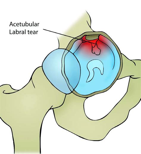
acetabular labral tear special tests|best imaging for labral tear : maker The gold standard for diagnosing an acetabular labral tear is considered to be direct visualization by arthroscopy. A medical physician may wish to utilize less invasive measures to make a diagnosis such as MR arthrography and or intra-articular hip injections with anesthetic. Our team specializes in autoclave repair services, catering to a spectrum of issues that could potentially disrupt your scientific endeavors. Our team of skilled technicians possesses the technical expertise to address issues with .
{plog:ftitle_list}
Use closed sterilization containers or a patented system such as the Flash-Guard system, so instruments are protected from contamination from autoclave to point-of-use. Such a system is easy-to-use, sterilization is fully .
The gold standard for diagnosing an acetabular labral tear is considered to be direct visualization by arthroscopy. A medical physician may wish to utilize less invasive measures to make a diagnosis such as MR arthrography and or intra-articular hip injections with anesthetic.Plain radiographs and computed tomography may show hip dysplasia, arthritis, and acetabul.
The gold standard for diagnosing an acetabular labral tear is considered to be direct visualization by arthroscopy. A medical physician may wish to utilize less invasive measures to make a diagnosis such as MR arthrography and or intra-articular hip injections with anesthetic.Plain radiographs and computed tomography may show hip dysplasia, arthritis, and acetabular cysts in patients with acetabular labrum tears, they are useful for excluding other types of hip pathology. MRI helps in diagnosing acetabular labral tears. A hip labral tear is a traumatic tear of the acetabular labrum, mostly common seen in acetabular dysplasia, that may lead to symptoms of internal snapping hip as well hip locking with hip range of motion. Diagnosis generally requires an MR arthrogram of the hip joint in question.A healthcare provider will diagnose a hip labral tear with a physical exam and some tests. They’ll examine your hip and ask you about your symptoms. Tell your provider when you first noticed pain and other symptoms, and if any activities, movements or positions make them worse.
Information regarding acetabular labral tears and their association to capsular laxity, femoral acetabular impingement (FAI), dysplasia of the acetabulum, and chondral lesions is emerging.
Imaging scans. A hip labral tear rarely occurs by itself. In most cases, other structures within the hip joint also have injuries. X-rays are excellent at visualizing bone. They can check for arthritis and for structural problems.Various clinical tests have been described, but little is known about their diagnostic sensitivity and specificity. We investigated the diagnostic validity of clinical tests . Background and purpose An acetabular labral tear is a diagnostic challenge.
This paper aims to assess the pathophysiology, diagnosis, and latest evidence-based treatment of acetabular labral tears. The acetabular labrum contributes to the stability of the hip. Labral tears may lead to significant pain and disability, although many are asymptomatic.
Clinical examination should consider lumbopelvic and extra-articular pathologies in addition to intra-articular pathologies when assessing for the source of symptoms and functional limitation. If a labral tear is suspected, further diagnostic testing may be indicated.
Arthroscopy remains the reference standard for diagnosing acetabular labral tears allowing for dynamic examination and assessing the extent of the tear [50, 52]. However, it is more invasive and may have limited access to posterior labral tears depending on portal positioning [52].The gold standard for diagnosing an acetabular labral tear is considered to be direct visualization by arthroscopy. A medical physician may wish to utilize less invasive measures to make a diagnosis such as MR arthrography and or intra-articular hip injections with anesthetic.
Plain radiographs and computed tomography may show hip dysplasia, arthritis, and acetabular cysts in patients with acetabular labrum tears, they are useful for excluding other types of hip pathology. MRI helps in diagnosing acetabular labral tears. A hip labral tear is a traumatic tear of the acetabular labrum, mostly common seen in acetabular dysplasia, that may lead to symptoms of internal snapping hip as well hip locking with hip range of motion. Diagnosis generally requires an MR arthrogram of the hip joint in question.
A healthcare provider will diagnose a hip labral tear with a physical exam and some tests. They’ll examine your hip and ask you about your symptoms. Tell your provider when you first noticed pain and other symptoms, and if any activities, movements or positions make them worse.Information regarding acetabular labral tears and their association to capsular laxity, femoral acetabular impingement (FAI), dysplasia of the acetabulum, and chondral lesions is emerging.
Imaging scans. A hip labral tear rarely occurs by itself. In most cases, other structures within the hip joint also have injuries. X-rays are excellent at visualizing bone. They can check for arthritis and for structural problems.Various clinical tests have been described, but little is known about their diagnostic sensitivity and specificity. We investigated the diagnostic validity of clinical tests . Background and purpose An acetabular labral tear is a diagnostic challenge. This paper aims to assess the pathophysiology, diagnosis, and latest evidence-based treatment of acetabular labral tears. The acetabular labrum contributes to the stability of the hip. Labral tears may lead to significant pain and disability, although many are asymptomatic.
Clinical examination should consider lumbopelvic and extra-articular pathologies in addition to intra-articular pathologies when assessing for the source of symptoms and functional limitation. If a labral tear is suspected, further diagnostic testing may be indicated.

right superior acetabular labral tear
labral tear mri images
HIP is a solid state process which applies heat and pressure simultaneously to objects in an autoclave via an inert gas in such a way as to eliminate internal voids and obtain desired .
acetabular labral tear special tests|best imaging for labral tear